Gallery
Brain Surgery
A) Cranipharyngioma.
Case 1:
Case 2:
Case 3:
B) Meningioma
Case 1:
Case 2:
Case 3:
C) Acoustic Neuroma
Case 1:
Case 2:
D) Colloid Cyst
E) Pituitary Adenoma
F) Vascular Surgery
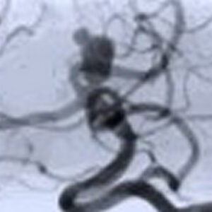
A preoperative cerebral arteriogram (A) shows a basilar tip aneurysm. A postoperative arteriogram, after aneurysm clipping via a superolateral orbital craniotomy, confirms successful clipping
G) Functional Surgery
Trigeminal Neuralgia
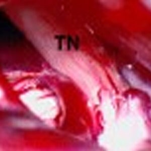
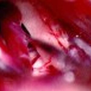
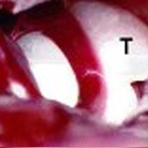
Intraoperative photographs show an arterial loop compressing the 5th cranial nerve (A; TN = trigeminal nerve), separation of the artery from the nerve (B), and placement of Teflon sponge (T) between the artery and the nerve (C).
H) Spine Surgery
Case 1:
Case 2:
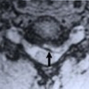
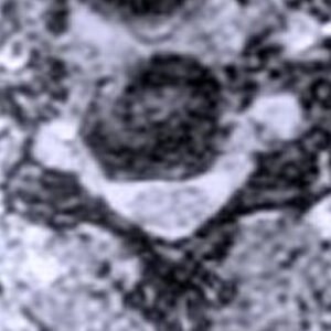
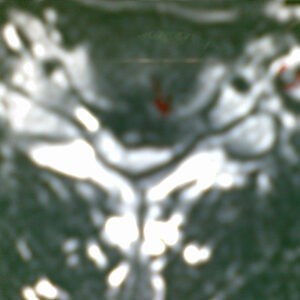
A preoperative MR scan, axial view (A), shows nerve root compression by herniated disc material at C5-6 (arrow). A postoperative MR scan, axial view (B) displays a widened nerve root canal after disc removal
Case 3:
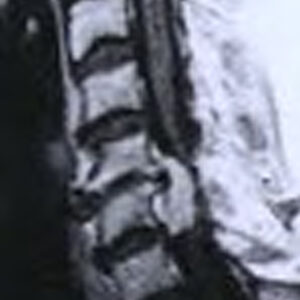
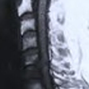
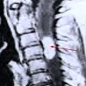
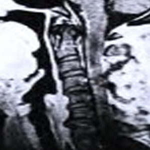
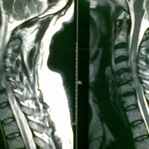
Preoperative MR scan, sagittal view (A), shows intraspinal tumour at C3-4(arrow). A postoperative MR scan, sagittal view (B) displays a complete excision
Case 4:
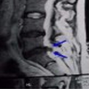
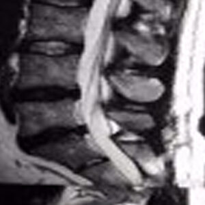
Preoperative MR scan, sagittal view (A), shows lumbar disc herniation. A postoperative MR scan, sagittal view (B) displays a complete decompression by Minimal Invasive technique.
Case 5:
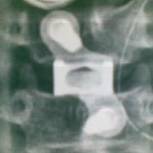
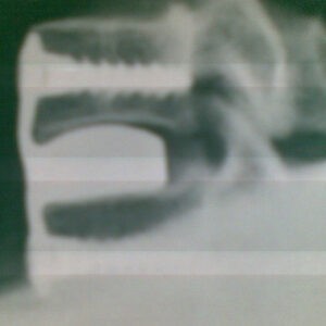
Preoperative image showing disc prolapse. Postoperative radiological images showing Plate Cage Fixation
Case 6:
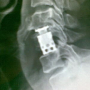
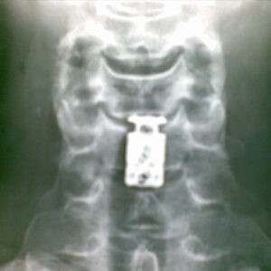
Preoperative image of traumatic fracture of cervical 5th vertebra. Postoperative radiological images showing C5 corpectomy and Fixation with Expandable Titanium Cage.
I) Paediatric Neurosurgery
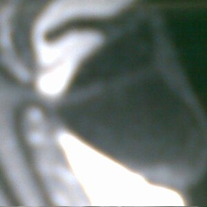
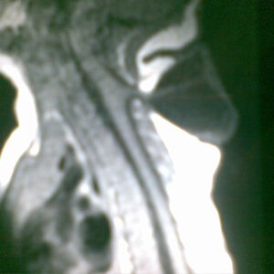
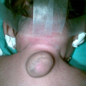
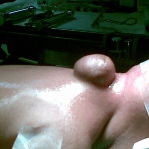
Preoperative and intraoperative pictures. This lesion was completely excised

